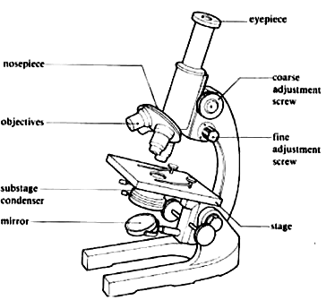
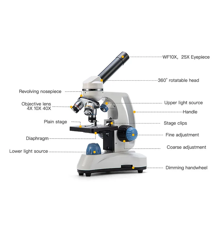
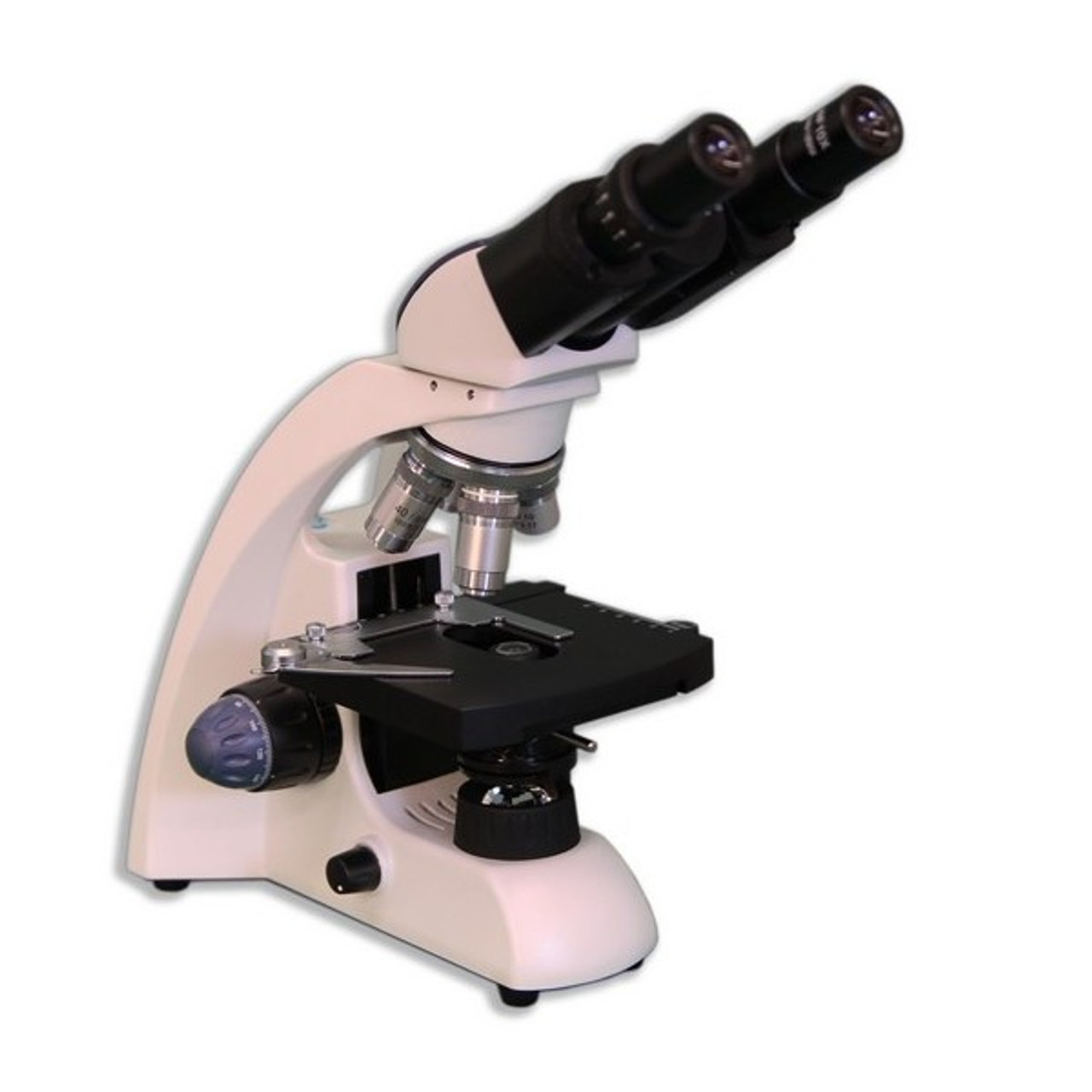
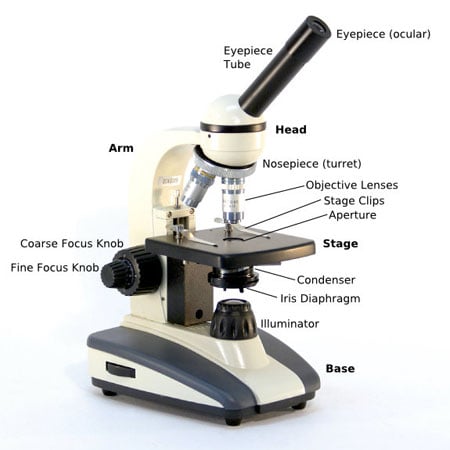
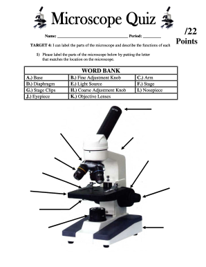
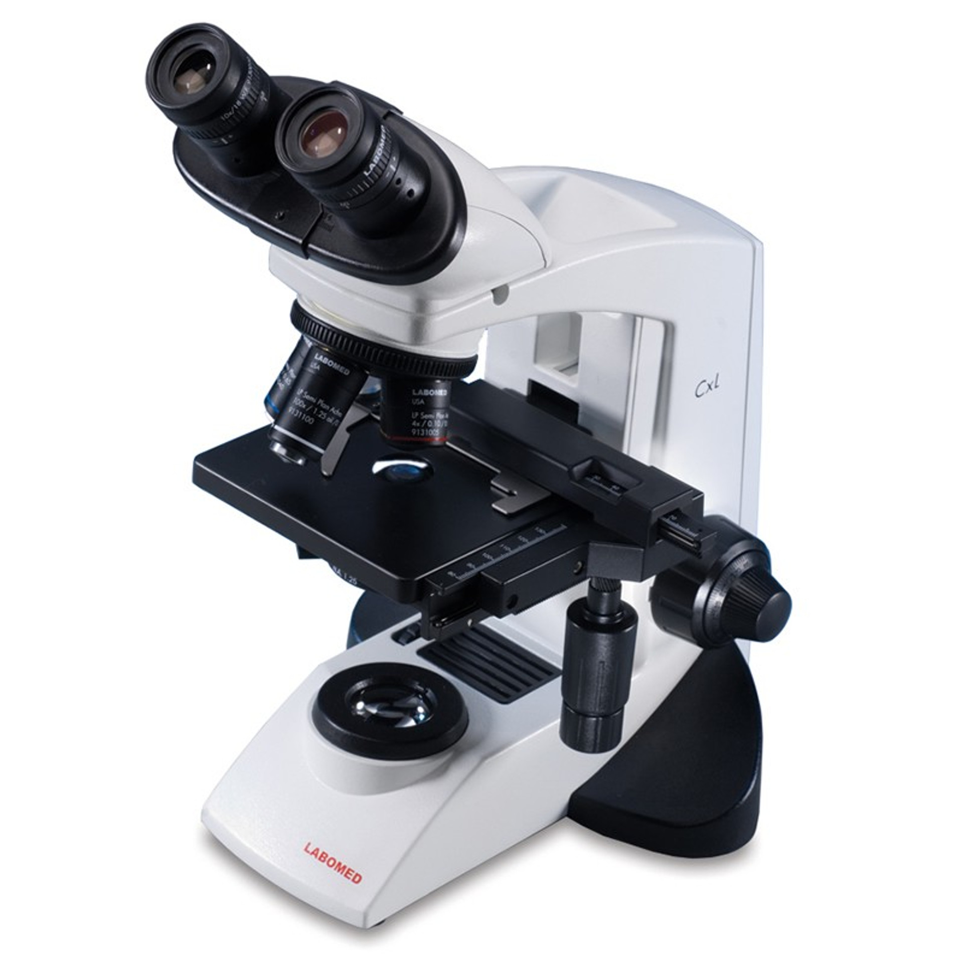
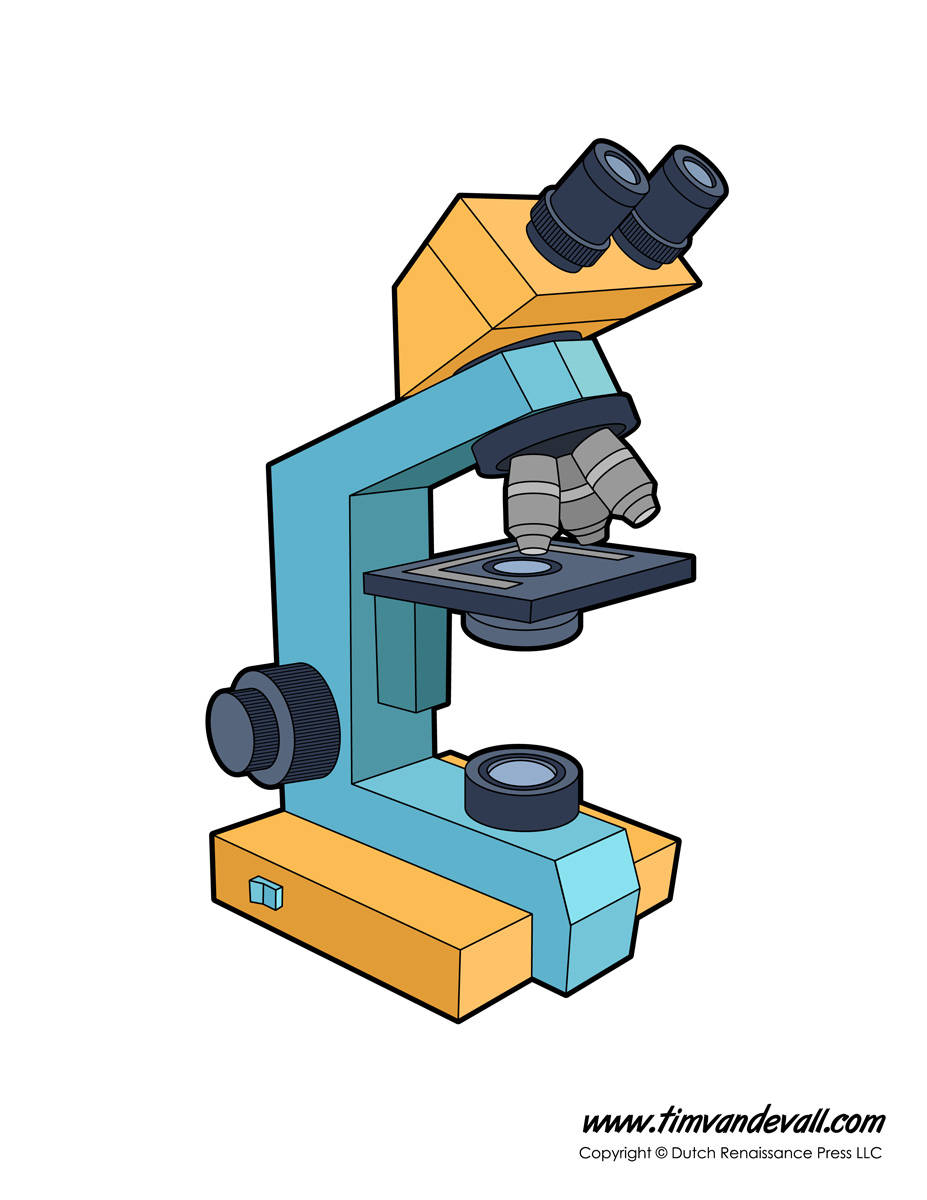
41 monocular microscope diagram with label
Microscope Types (with labeled diagrams) and Functions Has a higher level of magnification - Typically up to 2000x. Is used in hospitals and forensic labs by scientists, biologists and researchers to study micro organisms. Compound microscope labeled diagram. Compound microscope functions: It finds great application in areas of pathology, pedology, forensics etc. microbenotes.com › parts-of-a-microscopeParts of a microscope with functions and labeled diagram Sep 17, 2022 · For monocular microscopes, they are none flexible. Objective lenses – These are the major lenses used for specimen visualization. They have a magnification power of 40x-100X. There are about 1- 4 objective lenses placed on one microscope, in that some are rare facing and others face forward. Each lens has its own magnification power.
Simple Microscope - Parts, Functions, Diagram and Labelling Parts of the optical parts are as follows: Mirror - A simple microscope has a plano-convex mirror and its primary function is to focus the surrounding light on the object being examined. Lens - The biconvex lens is placed above the stage and its function is to magnify the size of the object being examined.
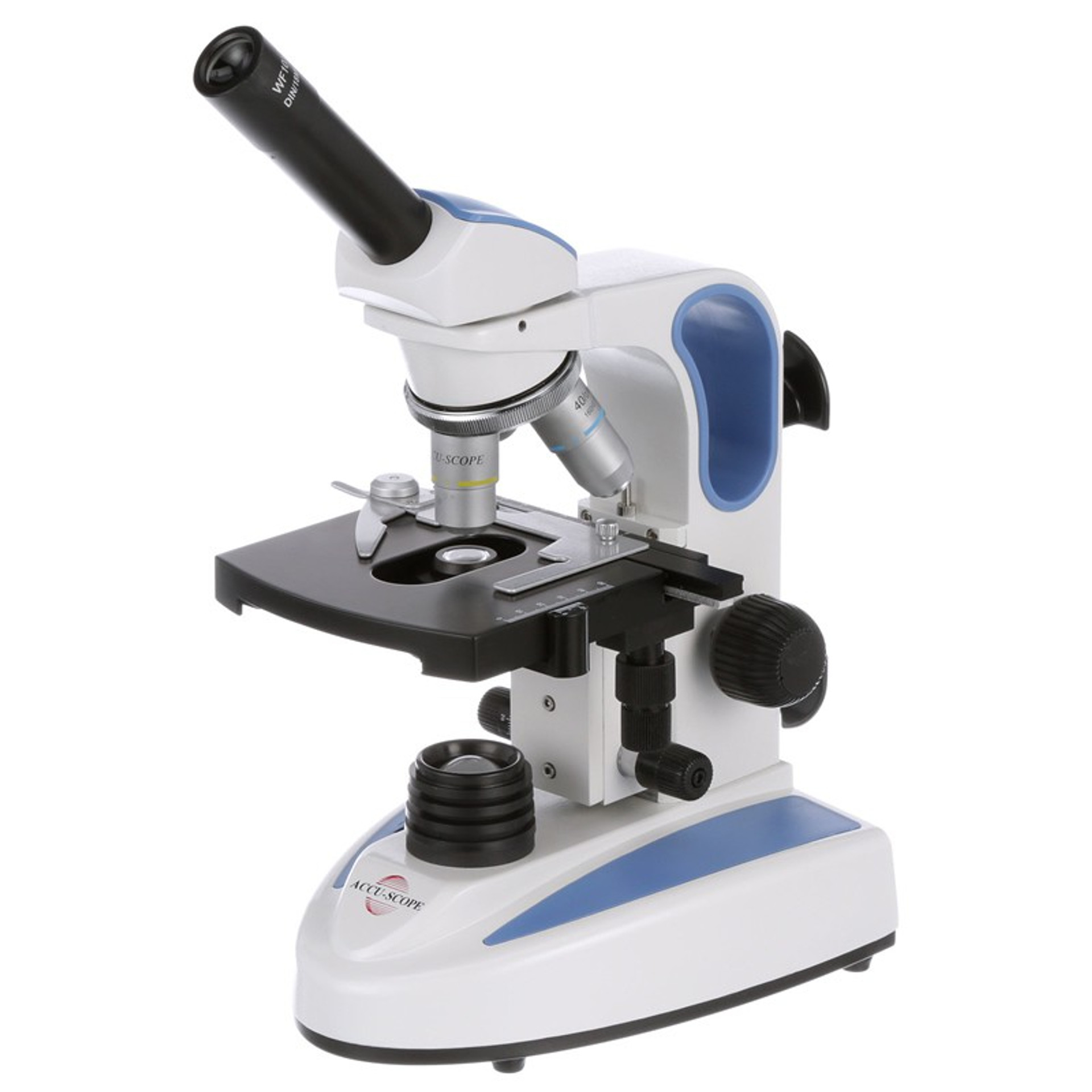
Monocular microscope diagram with label
Lifestyle | Daily Life | News | The Sydney Morning Herald The latest Lifestyle | Daily Life news, tips, opinion and advice from The Sydney Morning Herald covering life and relationships, beauty, fashion, health & wellbeing Parts of a microscope with functions and labeled diagram 17/09/2022 · For monocular microscopes, they are none flexible. Objective lenses – These are the major lenses used for specimen visualization. They have a magnification power of 40x-100X. There are about 1- 4 objective lenses placed on one microscope, in that some are rare facing and others face forward. Each lens has its own magnification power. Full Members | Institute Of Infectious Disease and Molecular … Full member Area of expertise Affiliation; Stefan Barth: Medical Biotechnology & Immunotherapy Research Unit: Chemical & Systems Biology, Department of Integrative Biomedical Sciences
Monocular microscope diagram with label. Microscopy: History, Types of Microscope, Diagram - Embibe A microscope known as a microscopy instrument is a device that magnifies pictures of tiny objects. Learn Microscopy history, diagrams, types, and parts. ... Diagram of Compound Microscope. ... It is the lens present at the top end of the metal tube. Monocular models have a single tube, whereas binocular models have two tubes, each with one ... › shows › fox-filesFox Files | Fox News Jan 31, 2022 · FOX FILES combines in-depth news reporting from a variety of Fox News on-air talent. The program will feature the breadth, power and journalism of rotating Fox News anchors, reporters and producers. Prolab Digital Compound Monocular Microscope, 2 MP Labels, markers and tapes; Microbiology; Mortars and pestles; pH measurement; Pipets; Plates and microplates; Scoops and spatulas; Stoppers; ... Prolab Digital Compound Monocular Microscope, 2 MP; Prolab Digital Compound Monocular Microscope, 2 MP. The built-in digital camera with a 2.0 megapixel resolution enhances the functionality of the ... BRESSER 5102000 Monocular Microscope Instruction Manual BRESSER 5102000 Monocular Microscope Instruction Manual CAUTION! To work with this microscope, sharp and pointed aids are being used. Please take care that this microscope and its accessories are stored at a place out of reach of children. Let children only work with this microscope under an adult's supervision! Keep packing material (plastic bags etc.) … Continue reading "BRESSER 5102000 ...
MIT - Massachusetts Institute of Technology a aa aaa aaaa aaacn aaah aaai aaas aab aabb aac aacc aace aachen aacom aacs aacsb aad aadvantage aae aaf aafp aag aah aai aaj aal aalborg aalib aaliyah aall aalto aam ... Microscope: Parts Of A Microscope With Functions And Labeled Diagram. The microscope has three basic components: the head, the base, and the arm. Head:Occasionally, the head is considered the body. It holds the optical components of the upper part of the microscope. Base:The microscope's base provides great support. It is also equipped with miniature illuminators. Simple Microscope - Diagram (Parts labelled), Principle, Formula and Uses Simple microscope is a magnification apparatus that uses a combination of double convex lens to form an enlarged, erect image of a specimen. The working principle of a simple microscope is that when a lens is held close to the eye, a virtual, magnified and erect image of a specimen is formed at the least possible distance from which a human eye ... Neuron under Microscope with Labeled Diagram - AnatomyLearner Neuron under microscope labelled diagram. Throughout this article, you got the different neurons labelled diagrams. Here, you will also find the diagrams of different neuron types under a microscope. The neuron diagram shows the different parts (axon, dendrites, and cell body) of the neurons.
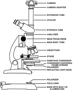
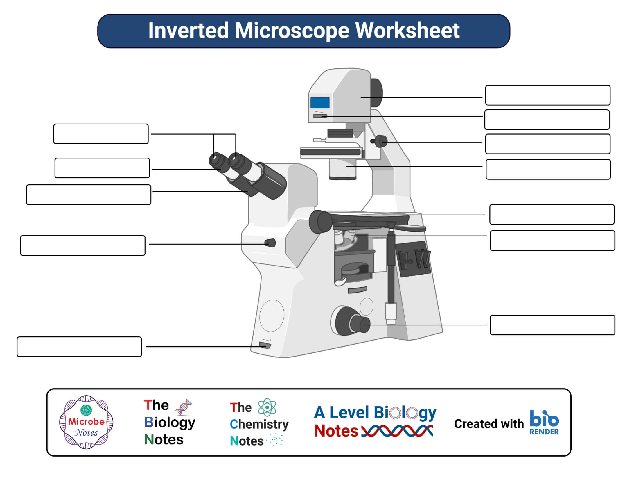
Electron Microscope Principle, Uses, Types and Images (Labeled Diagram ... Ans: A light microscope has a low resolving power (0.25µm to 0.3µm) while the electron microscope has a resolution power about 250 times higher than the light microscope at about 0.001µm. Similarly, a light microscope has a magnification of 500X to 1500x while the electron microscope has a much higher magnification of 100,000X to 300,000X. Compound Microscope - Diagram (Parts labelled), Principle and Uses See: Labeled Diagram showing differences between compound and simple microscope parts Structural Components. The three structural components include. 1. Head. This is the upper part of the microscope that houses the optical parts. 2. Arm . This part connects the head with the base and provides stability to the microscope. Givenchy official site Discover all the collections by Givenchy for women, men & kids and browse the maison's history and heritage Parts of a microscope with functions and labeled diagram (2022) Microscope Definition; Structural parts of a microscope and their functions. Figure: Diagram of parts of a microscope; Optical parts of a microscope and their functions; Parts of a Microscope Revision Questions (FAQs) Microscope Parts Worksheets. 1. Light Microscope Free Worksheet; 2. Inverted Microscope Free Worksheet; 3.
Compound Microscope- Definition, Labeled Diagram, Principle, Parts, Uses The optical microscope often referred to as the light microscope, is a type of microscope that uses visible light and a system of lenses to magnify images of small subjects. There are two basic types of optical microscopes: Simple microscopes. Compound microscopes. The term "compound" in compound microscopes refers to the microscope having ...
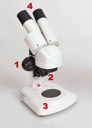
rsscience.com › stereo-microscopeParts of Stereo Microscope (Dissecting microscope) – labeled ... Labeled part diagram of a stereo microscope Major structural parts of a stereo microscope. There are three major structural parts of a stereo microscope. The viewing Head includes the upper part of the microscope, which houses the most critical optical components, including the eyepiece, objective lens, and light source of the microscope.
Fox Files | Fox News 31/01/2022 · FOX FILES combines in-depth news reporting from a variety of Fox News on-air talent. The program will feature the breadth, power and journalism of rotating Fox News anchors, reporters and producers.
Diagram of a Compound Microscope - Biology Discussion ADVERTISEMENTS: In this article we will discuss about:- 1. Essential Parts of Compound Microscope 2. Magnification of the Image of the Object by Compound Microscope 3. Resolution Power 4. Method for Studying Microbes 5. Measurement of the Size of Objects. Essential Parts of Compound Microscope: The essential parts of usually used monocular compound …
Parts of the Microscope with Labeling (also Free Printouts) 5. Knobs (fine and coarse) By adjusting the knob, you can adjust the focus of the microscope. The majority of the microscope models today have the knobs mounted on the same part of the device. Image 5: The circled parts of the microscope are the fine and coarse adjustment knobs. Picture Source: bp.blogspot.com.
Parts of Stereo Microscope (Dissecting microscope) – labeled diagram ... Unlike a compound microscope that offers a flat image, stereo microscopes give the viewer a 3-dimensional image that you can see the texture of a larger specimen. [In this image] Examples of Stereo & Dissecting microscopes. Major microscope brands (Zeiss, Olympus, Nikon, Amscope, Omano, Leica …) all produce stereomicroscopes.
Microscope Parts, Function, & Labeled Diagram - slidingmotion Diaphragm. The diaphragm is also called as iris. This iris situates below the stage of the microscope. The function of the diaphragm is to control the amount of light that focuses on the specimen. This diaphragm can adjust the amount of light and intensity of light that falls on the specimen. In some standard and high-quality microscopes, this ...
Components of a microscope with features and labeled diagram There are three structural elements of the microscope i.e. head, base, and arm. Head - That is also called the physique. It carries the optical elements within the higher a part of the microscope. Base - It acts as microscopes help. It additionally carries microscopic illuminators.
Microscope, Microscope Parts, Labeled Diagram, and Functions Revolving Nosepiece or Turret: Turret is the part of the microscope that holds two or multiple objective lenses and helps to rotate objective lenses and also helps to easily change power. Objective Lenses: Three are 3 or 4 objective lenses on a microscope. The objective lenses almost always consist of 4x, 10x, 40x and 100x powers. The most common eyepiece lens is 10x and when it coupled with ...
Microscope: Types of Microscope, Parts, Uses, Diagram - Embibe There microscope anatomy includes three structural parts, i.e. head, base, and arm. Head - This is also known as the body; it carries the optical parts in the upper part of the microscope.. Base - It acts as microscopes support.It also carries microscopic illuminators. Arms - The microscope arm connects the base and the head and the eyepiece tube to the microscope base.
Stanford University UNK the , . of and in " a to was is ) ( for as on by he with 's that at from his it an were are which this also be has or : had first one their its new after but who not they have
Electron Microscope- Definition, Principle, Types, Uses, Labeled Diagram There are two types of electron microscopes, with different operating styles: 1. Transmission Electron Microscope (TEM) The transmission electron microscope is used to view thin specimens through which electrons can pass generating a projection image. The TEM is analogous in many ways to the conventional (compound) light microscope.
dx.doi.orgResolve a DOI Name Type or paste a DOI name into the text box. Click Go. Your browser will take you to a Web page (URL) associated with that DOI name. Send questions or comments to doi ...
Simple Squamous Epithelium under a Microscope with a Labeled Diagram ... Here the artery labeled diagram shows the tunica intima that consists of endothelium, basal lamina, subendothelium connective tissue, and internal elastic lamina. You will find the endoplasmic reticulum and mitochondria in the cytoplasm of the endothelium cell under the electron microscope.
› intGivenchy official site Discover all the collections by Givenchy for women, men & kids and browse the maison's history and heritage
› lifestyleLifestyle | Daily Life | News | The Sydney Morning Herald The latest Lifestyle | Daily Life news, tips, opinion and advice from The Sydney Morning Herald covering life and relationships, beauty, fashion, health & wellbeing
Resolve a DOI Name Type or paste a DOI name into the text box. Click Go. Your browser will take you to a Web page (URL) associated with that DOI name. Send questions or comments to doi ...
› microscope › compoundDiagram of a Compound Microscope - Biology Discussion ADVERTISEMENTS: In this article we will discuss about:- 1. Essential Parts of Compound Microscope 2. Magnification of the Image of the Object by Compound Microscope 3. Resolution Power 4. Method for Studying Microbes 5. Measurement of the Size of Objects. Essential Parts of Compound Microscope: The essential parts of usually used monocular compound microscope (Fig. […]
Full Members | Institute Of Infectious Disease and Molecular … Full member Area of expertise Affiliation; Stefan Barth: Medical Biotechnology & Immunotherapy Research Unit: Chemical & Systems Biology, Department of Integrative Biomedical Sciences
Parts of a microscope with functions and labeled diagram 17/09/2022 · For monocular microscopes, they are none flexible. Objective lenses – These are the major lenses used for specimen visualization. They have a magnification power of 40x-100X. There are about 1- 4 objective lenses placed on one microscope, in that some are rare facing and others face forward. Each lens has its own magnification power.
Lifestyle | Daily Life | News | The Sydney Morning Herald The latest Lifestyle | Daily Life news, tips, opinion and advice from The Sydney Morning Herald covering life and relationships, beauty, fashion, health & wellbeing

Happybuy Monocular Compound Microscope 40X-2500X Magnification Digital Compound Microscope WF10X & WF25X Eyepieces Compound Light Microscope Tungsten ...

SWIFT Microscopes for Kids,Monocular Microscopes for Children Beginners with Educational Science Microscope Kits, Slides and LED Light














Post a Comment for "41 monocular microscope diagram with label"