43 light microscope with labels
Parts of a Microscope Labeling Activity - Storyboard That In this activity, students will create a poster of a microscope with labeled parts. Students will identify and describe the microscope parts and functions. This is an awesome activity to complete at the beginning of either the school year or the unit on basic cells. ... Provides light to illuminate the specimen, sometimes a mirror is also used ... › products › microscopeLAS X Industry Microscope software for Industry | Products ... The software can handle multiple users who have different levels of microscope skills and diverse tasks to accomplish. Profiles according to user’s skills. The LAS X software enables you to create profiles according to the skills and tasks of individual users – from microscopy beginner to expert. It helps you to get reliable results.
Fluorescence Microscopy - Explanation and Labelled Images A fluorescence microscope is used to study organic and inorganic samples. Fluorescence microscopy uses fluorescence and phosphorescence to examine the structural organization, spatial distribution of samples. It is particularly used to study samples that are complex and cannot be examined under conventional transmitted-light microscope.
Light microscope with labels
Label the Light Microscope - Labelled diagram - Wordwall Drag and drop the pins to their correct place on the image.. Eyepiece, Light Source, Base, Stage, Stage Clips, Fine Focus, Coarse Focus, Arm, Objective Lens. Compound Light Microscope: Everything You Need to Know A fluorescence microscope, also called a confocal microscope, is a kind of biological microscope that operates by using different light colors and wavelengths over-dyed specimen samples in order for the dye to interact with the light, after which the resulting image is scanned. Microscope Objective Lens | Products | Leica Microsystems The objective lens is a critical part of the microscope optics. The microscope objective is positioned near the sample, specimen, or object being observed. It has a very important role in imaging, as it forms the first magnified image of the sample. The numerical aperture (NA) of the objective indicates its ability to gather light and largely determines the microscope’s …
Light microscope with labels. › microscopy › enZEISS Elyra 7 with Lattice SIM² Super-Resolution Microscope Lattice SIM² comes with outstanding out-of-focus light suppression, giving you the sharpest sectioning in widefield microscopy even for highly scattering samples. SIM² image reconstruction robustly reconstructs all structured-illumination-based acquisition data of your Elyra 7 – with minimal artefacts – for living and fixed samples. What is Electron Microscopy? - UMASS Medical School The transmission electron microscope is used to view thin specimens (tissue sections, molecules, etc) through which electrons can pass generating a projection image. The TEM is analogous in many ways to the conventional (compound) light microscope. TEM is used, among other things, to image the interior of cells (in thin sections), the structure of protein molecules … Light Labs distributes PCR... Welcome to Light Labs. Since 2002, Light Labs has distributed high quality laboratory consumables and equipment, including MultiMax Barrier tips, PCR tubes and strip tubes, PCR plates, and much more. With an emphasis on customer service, we have successfully served the research marketplace with a wide array of laboratory goods. Compound Microscope Labeled Diagram | Quizlet Part that supports the microscope. Stage. Supports the slide or specimen. Coarse adjustment Knob. sed to focus when using the low power objective lenses. Fine Adjustment Knob. Used to focus the image on high power to view image in more detail. Revolving nose piece. The revolving piece on which the lenses are attached.
Label a microscope - Teaching resources - Wordwall Label a microscope - Label the Light Microscope - Label the Light Microscope - Microscope slide - label the parts - Year 7 A Microscope. › microscopy › enZEISS Lattice Lightsheet 7 The importance of gentle light sheet imaging at high resolution cannot be overestimated for the study of subcellular processes. With Lattice Lightsheet 7, ZEISS makes access to the benefits of this advanced technology amazingly simple. Sperm Under Microscope with Labeled Diagram - AnatomyLearner Under the light microscope, the sperm consists of two main portions - the head and the tail. But, the electron microscope shows four different parts in the tail of spermatozoa. ... So, this article provides the details structural features of sperm under the light microscope. All the labeled diagrams might help you identify the sperms from ... rsscience.com › stereo-microscopeParts of Stereo Microscope (Dissecting microscope) – labeled ... A stereo microscope allows you to see the surface of specimens with a 3-dimensional view. Under a stereo microscope, you can see the metallic texture and colors of the mosquito’s compound eyes. In contrast, the light has to pass through the specimen to form the image under a compound microscope.
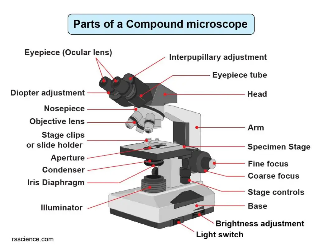
Compound Light Microscope Labelling Quiz - PurposeGames.com This is an online quiz called Compound Light Microscope Labelling There is a printable worksheet available for download here so you can take the quiz with pen and paper. Your Skills & Rank Total Points 0 Get started! Today's Rank -- 0 Today 's Points One of us! Game Points 15 You need to get 100% to score the 15 points available Actions Addgene: Using a Light Microscope Protocol Figure 1: Diagram of a compound light microscope with labels. Created with BioRender.com. Base Light Source Condenser and Diaphragm Stage Objective Lenses Focus Knobs (Fine and Coarse) Nosepiece Arm Ocular Lens (Eyepiece) The route the light follows from the source to your eyes is called the light path. Parts of a microscope with functions and labeled diagram - Microbe Notes Microscopic illuminator - This is the microscopes light source, located at the base. It is used instead of a mirror. It captures light from an external source of a low voltage of about 100v. Condenser - These are lenses that are used to collect and focus light from the illuminator into the specimen. ZEISS Lattice Lightsheet 7 Gentle illumination is crucial for imaging mitosis as this process is extremely delicate and light sensitive. To prevent replication of damaged DNA, cells arrest mitosis as soon as there is any damage from excitation light. The gentleness of Lattice Lightsheet 7 imaging and an extremely stable system is required for imaging mitotic events over ...
Compound Microscope Parts - Labeled Diagram and their Functions The eyepiece (or ocular lens) is the lens part at the top of a microscope that the viewer looks through. The standard eyepiece has a magnification of 10x. You may exchange with an optional eyepiece ranging from 5x - 30x. [In this figure] The structure inside an eyepiece. The current design of the eyepiece is no longer a single convex lens.
Compound Microscope Parts, Functions, and Labeled Diagram Compound Microscope Definitions for Labels. Eyepiece (ocular lens) with or without Pointer: The part that is looked through at the top of the compound microscope. Eyepieces typically have a magnification between 5x & 30x. Monocular or Binocular Head: Structural support that holds & connects the eyepieces to the objective lenses.
Microscope Labels Flashcards | Quizlet condenser. iris diaphragm. stage. stage clip. on off switch. light intensity knob. fine focus knob. course focus knob. mechanical stage adjustment knobs.
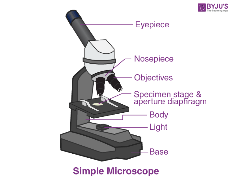
Simple Microscope - Diagram (Parts labelled), Principle, Formula ... Feb 23, 2022 ... Dating back to the 14th century, simple microscope is the most basic of the various microscopes available. It is a type of optical ...
proscitech.com.auProSciTech Laboratory supplies and Lab equipment for Histology, Pathology, Light Microscopy, Electron Microscopy and specialist researchers.
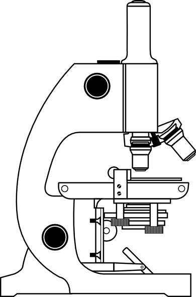
Microscope With Labels Clip Art at Clker.com PEOPLE GOT HERE BY SEARCHING: diagrams of the microscope · light microscope and label · the compound microscope drawing · diagram of microscope with labelling ...
LAS X Industry Microscope software for Industry | Products The software can handle multiple users who have different levels of microscope skills and diverse tasks to accomplish. Profiles according to user’s skills. The LAS X software enables you to create profiles according to the skills and tasks of individual users – from microscopy beginner to expert. It helps you to get reliable results.
Labeling the Parts of the Microscope - Pinterest Jan 13, 2016 - Free worksheets for labeling parts of the microscope including a worksheet that is ... Microscope, Assessment, Labels, Science, Education,.
Compound Microscope - Diagram (Parts labelled), Principle and Uses Also called as binocular microscope or compound light microscope, it is a remarkable magnification tool that employs a combination of lenses to magnify the image of a sample that is not visible to the naked eye. Compound microscopes find most use in cases where the magnification required is of the higher order (40 - 1000x).
ProSciTech Laboratory supplies and Lab equipment for Histology, Pathology, Light Microscopy, Electron Microscopy and specialist researchers.
Microscope Labeling Game - PurposeGames.com This is an online quiz called Microscope Labeling Game There is a printable worksheet available for download here so you can take the quiz with pen and paper. This quiz has tags. Click on the tags below to find other quizzes on the same subject. Science microsope Your Skills & Rank Total Points 0 Get started! Today's Rank -- 0 's Points 15 Actions
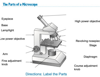
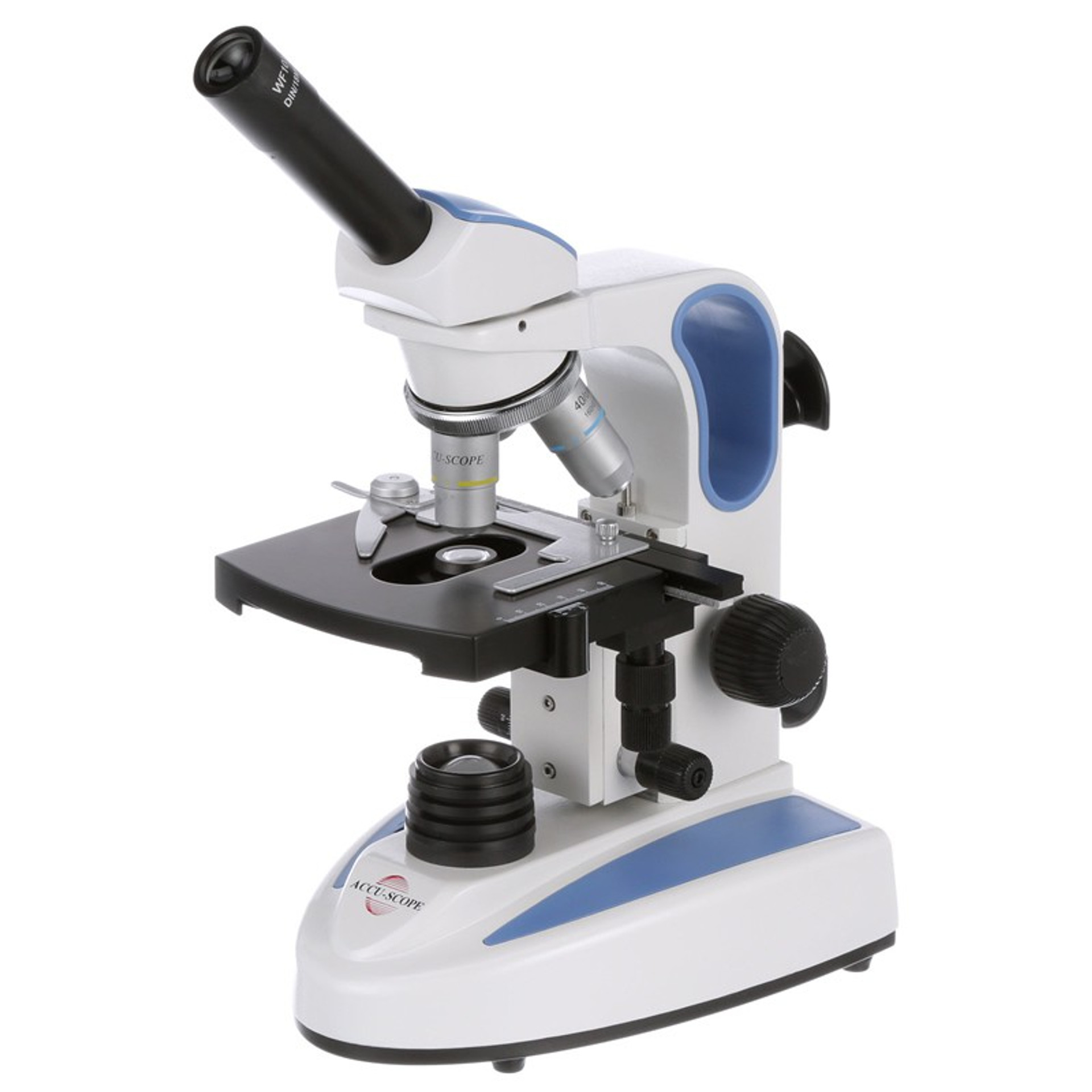
Microscope Labeling - The Biology Corner Students label the parts of the microscope in this photo of a basic laboratory light microscope. Can be used for practice or as a quiz. Name_____ Microscope Labeling . Microscope Use: 15. When focusing a specimen, you should always start with the _____ objective.
Label the light microscope | Teaching Resources Label the light microscope. Subject: Biology. Age range: 11-14. Resource type: Worksheet/Activity (no rating) 0 reviews. Science Resources. 4 1 reviews. Hi there, I upload a mixture of resources with a focus on assessment for learning and literacy within science, as well as general resources that I have used in the classroom and think would be ...
Labs distributes PCR... Welcome to Light Labs. Since 2002, Light Labs has distributed high quality laboratory consumables and equipment, including MultiMax Barrier tips, PCR tubes and strip tubes, PCR plates, and much more. With an emphasis on customer service, we have successfully served the research marketplace with a wide array of laboratory goods.
Labeling the Parts of the Microscope | Microscope World Resources Labeling the Parts of the Microscope This activity has been designed for use in homes and schools. Each microscope layout (both blank and the version with answers) are available as PDF downloads. You can view a more in-depth review of each part of the microscope here. Download the Label the Parts of the Microscope PDF printable version here.
Microscope Parts and Functions Body tube (Head): The body tube connects the eyepiece to the objective lenses. Arm: The arm connects the body tube to the base of the microscope. Coarse adjustment: Brings the specimen into general focus. Fine adjustment: Fine tunes the focus and increases the detail of the specimen. Nosepiece: A rotating turret that houses the objective lenses.
A Study of the Microscope and its Functions With a Labeled Diagram ... These labeled microscope diagrams and the functions of its various parts, attempt to simplify the microscope for you. However, as the saying goes, 'practice makes perfect', here is a blank compound microscope diagram and blank electron microscope diagram to label.
Labelled Diagram Of A Light Microscope | Products & Suppliers There are many applications for solar simulators and light simulators. Some products are designed to test coatings and paints, paper and labels, optical ...
Parts of Stereo Microscope (Dissecting microscope) – labeled … A stereo microscope allows you to see the surface of specimens with a 3-dimensional view. Under a stereo microscope, you can see the metallic texture and colors of the mosquito’s compound eyes. In contrast, the light has to pass through the specimen to form the image under a compound microscope. In this case, the region of compound eyes is ...
Light Microscope: Functions, Parts and How to Use It To use a light microscope, you can follow the steps below carefully. Start with a low lens and a clean slide. The microscope stage should be lowered as low as possible. Center the slide so that the specimen is under the objective lens. Use the coarse adjustment knob to get a general focus. Then slowly move up the stage until focus is achieved.
Microscope Labeling - The Biology Corner 1) Start with scanning (the shortest objective) and only use the COARSE knob . Once it is focused… 2) Switch to low power (medium) and only use the COARSE knob . You may need to recenter your slide. Once it is focused.. 3) Switch to high power (long objective).
Microscope With Labels clip art - Pinterest Jul 3, 2012 - Download Clker's Microscope With Labels clip art and related ... Optical Microscope, Microscopes, Focus Light, Industrial Machine, Things.
ZEISS Elyra 7 with Lattice SIM² Super-Resolution Microscope Despite being a structured illumination-based microscope, Elyra 7 Lattice SIM² as well as SIM² Apotome also provide you with super-resolution and high-quality sectioning in thick or scattering samples. The combination of robust illumination patterns and excellent image reconstruction technology enabled us to image throughout an entire murine brain section of ~80 µm thickness …
Light microscopes - Cell structure - Edexcel - BBC Bitesize Microscopes are used to produce magnified images. There are two main types of microscope: light microscopes are used to study living cells and for regular use when relatively low magnification and...
Amazon.com : LCD Digital Microscope,ANNLOV 4.3 inch … 05/02/2020 · This item LCD Digital Microscope,ANNLOV 4.3 inch Handheld USB Microscope 50X-1000X Magnification Coin Microscope Video Camera with 8 Adjustable LED Lights for Adults PCB Soldering Kids Outside Use ANNLOV 7" LCD Digital Microscope with 32GB TF Card 1200X Maginfication 1080P Coin Microscope with Wired Remote,12MP Ultra-Precise Focusing Video …
Barcode Labels and Tags | Zebra 8000T Light-Weight Tag: Tyvek® Olefin Tag: Thermal Transfer: Light-weight olefin label that provides tear resistance and durability. Ideal for lawn tags and garment tags. 8000T Light-Weight Tag. 6000 Wax; 6100 Wax/Resin. 8000T Tuff Tag: V-Max® Polyolefin Tag: Thermal Transfer: Provides good cross tear resistance and outdoor use for up to 1 ...
Light Microscope- Definition, Principle, Types, Parts, Labeled Diagram ... A light microscope is a biology laboratory instrument or tool, that uses visible light to detect and magnify very small objects and enlarge them. They use lenses to focus light on the specimen, magnifying it thus producing an image. The specimen is normally placed close to the microscopic lens.
Parts of the Microscope with Labeling (also Free Printouts) Parts of the Microscope with Labeling (also Free Printouts) By Editorial Team March 7, 2022 A microscope is one of the invaluable tools in the laboratory setting. It is used to observe things that cannot be seen by the naked eye. Table of Contents 1. Eyepiece 2. Body tube/Head 3. Turret/Nose piece 4. Objective lenses 5. Knobs (fine and coarse) 6.
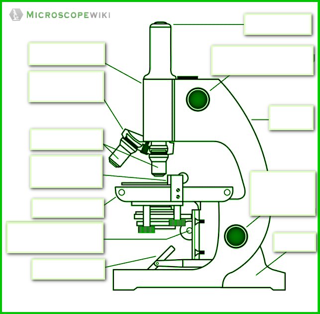
Label the microscope — Science Learning Hub All microscopes share features in common. In this interactive, you can label the different parts of a microscope. Use this with the Microscope parts activity to help students identify and label the main parts of a microscope and then describe their functions. Drag and drop the text labels onto the microscope diagram.
› products › microscopeMicroscope Objective Lens | Products | Leica Microsystems The objective lens is a critical part of the microscope optics. The microscope objective is positioned near the sample, specimen, or object being observed. It has a very important role in imaging, as it forms the first magnified image of the sample. The numerical aperture (NA) of the objective indicates its ability to gather light and largely determines the microscope’s resolution, the ...
Microscope Types (with labeled diagrams) and Functions This is an advanced microscope that has specific application in viewing, observing and measuring the optical thickness and phase of completely transparent specimens and objects. A tiny interferometer is used and a specimen is placed on beam path of it. This path is split and then rejoined to create two superimposed images of the specimen in focus.
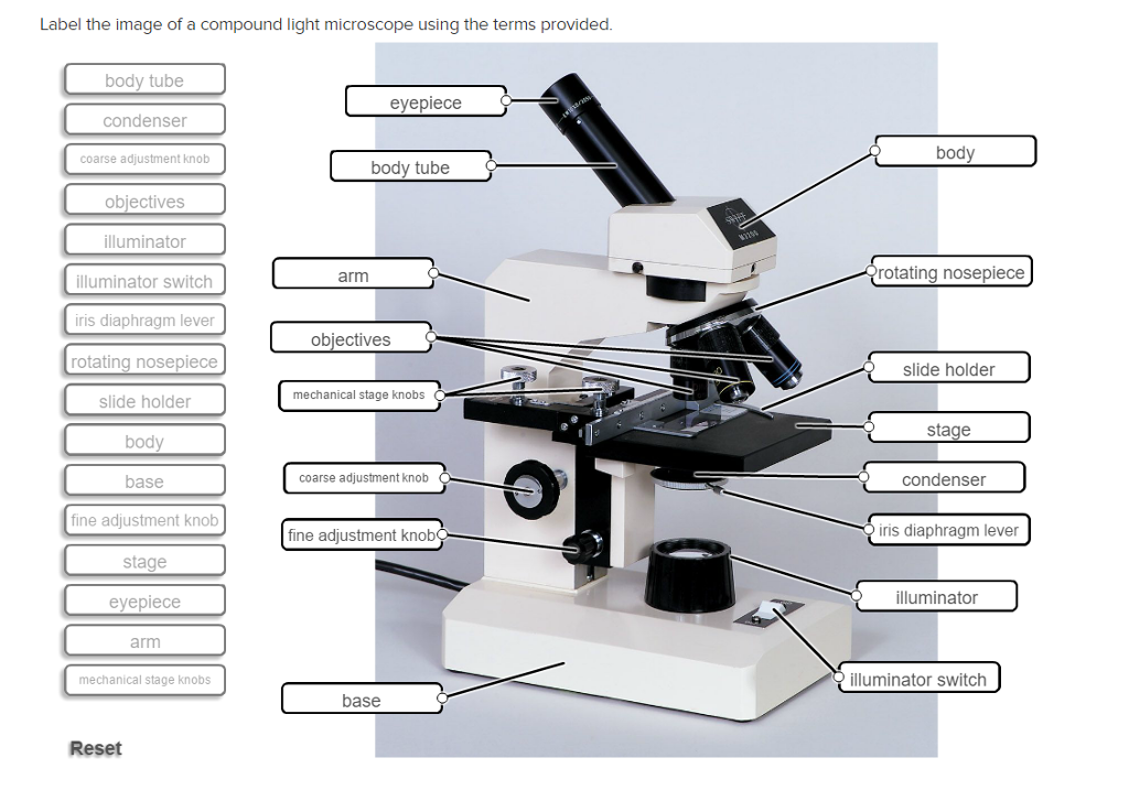
Solved Label the image of a compound light microscope using - Chegg Expert Answer. 100% (17 ratings) Transcribed image text: Label the image of a compound light microscope using the terms provided.
Microscope Labeled Pictures, Images and Stock Photos Diagram of the process of photosynthesis, showing the light reactions and the Calvin cycle. photosynthesis by absorbing water, light from the sun, and carbon dioxide from the atmosphere and converting it to sugars and oxygen. Light reactions occur in the thylakoid. Calvin Cycle occurs in the stoma. microscope labeled stock illustrations
Microscope Objective Lens | Products | Leica Microsystems The objective lens is a critical part of the microscope optics. The microscope objective is positioned near the sample, specimen, or object being observed. It has a very important role in imaging, as it forms the first magnified image of the sample. The numerical aperture (NA) of the objective indicates its ability to gather light and largely determines the microscope’s …
Compound Light Microscope: Everything You Need to Know A fluorescence microscope, also called a confocal microscope, is a kind of biological microscope that operates by using different light colors and wavelengths over-dyed specimen samples in order for the dye to interact with the light, after which the resulting image is scanned.
Label the Light Microscope - Labelled diagram - Wordwall Drag and drop the pins to their correct place on the image.. Eyepiece, Light Source, Base, Stage, Stage Clips, Fine Focus, Coarse Focus, Arm, Objective Lens.









.png)


Post a Comment for "43 light microscope with labels"